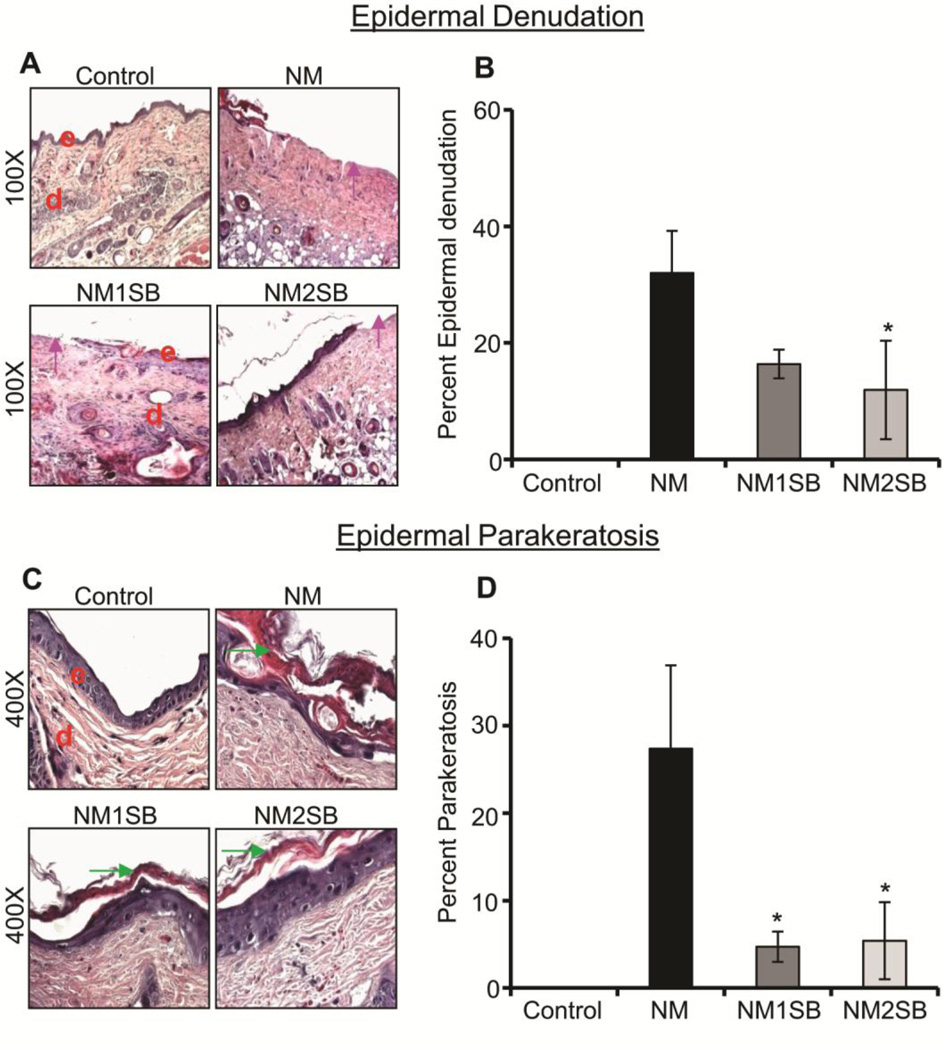
Figure 2. Effect of silibinin treatment on NM-induced epidermal denudation and parakeratosis.
H&E stained dorsal skin sections were analyzed for epidermal denudation and epidermal parakeratosis at 72h. (A) and (C) are representative pictures of epidermal denudation and epidermal parakeratosis, respectively. (B) and (D) Depict quantitative data for epidermal denudation and epidermal parakeratosis, respectively. Data presented are the mean ±SEM from 4–5 animals. *, p<0.05 compared to NM exposed group. e, epidermis; d, dermis; pink arrow, epidermal denuding; green arrow, epidermal parakeratosis.

