
Figure 3.
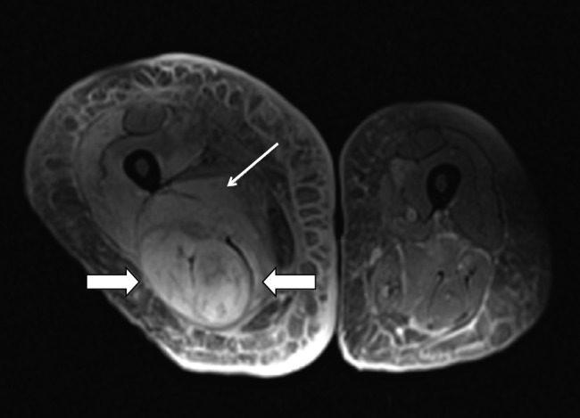
Imaging studies. Proton density fat-saturated MRI sequence demonstrates an increased signal within the semimembranosus and biceps femoris musculature (large arrows) and the adductor magnus muscle (thin arrow) consistent with edema due to early muscle infarction. MRI was performed in the 103 cases included. The findings identified included T2 hyperintensity (86; 76.8%), T1 hypointensity (7; 6.3%), T1 isointensity (8; 7.1%), and T1 hyperintensity (2; 1.8%). No MRI was performed in 23 cases (18.3%).