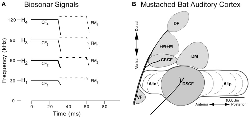
Figure 2.
Echolocation and the functional organization of the mustached bat auditory cortex. (A) H1−4 refer to harmonics 1 through 4 of the echolocation pulse or echo. Note the constant frequency (CF) and frequency-modulated (FM) components present in the pulse and echo. (B) Lateral view of the mustached bat auditory cortex showing the location of the DSCF area (shown in gray) as defined based on its role in computing biosonar signals. Anatomical landmarks (blood vessels shown by thick lines) and tuning properties of neuronal responses were used to identify the Doppler-shifted constant frequency (DSCF), anterior primary auditory (A1a), posterior primary auditory (A1p), dorsomedial (DM), CF/CF, FM-FM, and dorsal fringe (DF) areas (adapted from Suga et al., 1983).

