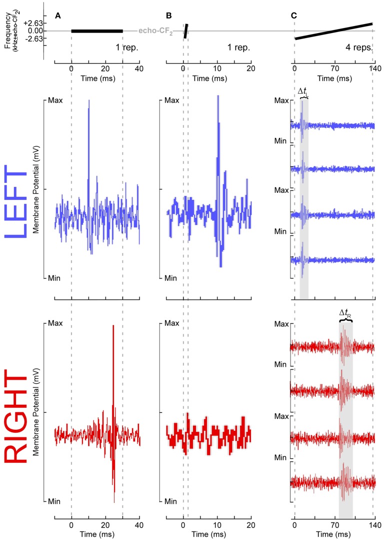
Figure 4.
Temporal response parameters of DSCF neurons as evidence for asymmetric sampling of time in mustached bats. All stimuli presented in the echo-CF2 range (57.5–60 kHz in P.p. rubiginosus) were paired at onset with a 30-ms CF tone-burst in the pulse-FM1 (23–28 kHz). Responses shown in (A–C) are from six different DSCF neurons, selected because they best illustrated a particular ASTIR-related concept. (A top): A 30-ms, constant-frequency tone presented at echo-CF2. (A middle): Voltage trace from a typical left hemispheric DSCF neuron in a male mustached bat following one presentation of a 30-ms CF tone-burst presented at the neuron's best frequency (BF) and best amplitude of excitation (BAE). This neuron is responding within 10 ms after stimulus onset. (A bottom): Voltage trace from a typical right hemisphere DSCF neuron in a male mustached bat following one presentation at BAE of a 30-ms CF tone-burst centered on the neuron's BF. This neuron is responding >20 ms after stimulus onset. Left DSCF neurons typically respond to tonal stimuli 3–5 ms before those on the right in male, but not female, bats (Washington and Kanwal, 2012). Assuming DSCF neurons conform to typical integrate-and-fire models, in male moreso than female bats, ASTIR takes the form of left DSCF neurons to integrating salient stimulus features and firing in less time than right DSCF neurons. (B top): A 1.31-ms, upward FM centered on echo-CF2, which has a modulation rate of 4 kHz/ms and a bandwidth of 5.25 kHz. (B middle): Voltage trace from a typical left DSCF neuron in a male bat following one presentation of a 5.25 kHz, 4 kHz/ms upward FM at BAE and centered on the neuron's BF. This neuron is responding within 10 ms after stimulus onset. (B bottom): Voltage trace from a typical right DSCF neuron in a male bat following presentation at BAE of a 5.25 kHz, 4 kHz/ms upward FM centered at the neuron's BF. This neuron is simply not responding. Relative to left DSCF neurons, right DSCF neurons are less responsive to shorter FM signals (Washington and Kanwal, 2012). This selectivity for longer sounds suggests right DSCF neurons have longer integration windows and are thus less likely to respond to such short sounds. Though this hemispheric difference is observed in both sexes, it is more pronounced in males. (C top): A 131-ms, upward FM centered at echo-CF2, which has a modulation rate of 0.04 kHz/ms and a bandwidth of 5.25 kHz. (C middle): Voltage traces from a typical left DSCF neuron in a male bat following four presentations at BAE of a 5.25 kHz, 0.04 kHz/ms upward FM centered on the neuron's BF. This neuron's four responses are time-locked and occur within the first 30 ms of the stimulus. (C bottom): Voltage traces from a typical right DSCF neuron in a male bat following four presentations at BAE of a 5.25 kHz, 0.04 kHz/ms upward FM centered at the neuron's BF. This neuron's four responses are not time-locked (i.e., tonic or burst firing) and occur after the first 70 ms of the stimulus. In both sexes, the maximum response duration of the left DSCF neuron (ΔtL) is less than that of the right DSCF neuron (ΔtR) (Washington and Kanwal, 2012). Since, in general, ΔtR > ΔtL, right DSCF neurons in general are less capable of processing precise temporal information than left DSCF neurons. Washington and Kanwal, unpublished data, reproduced with the permission of Jagmeet S. Kanwal, PhD.

