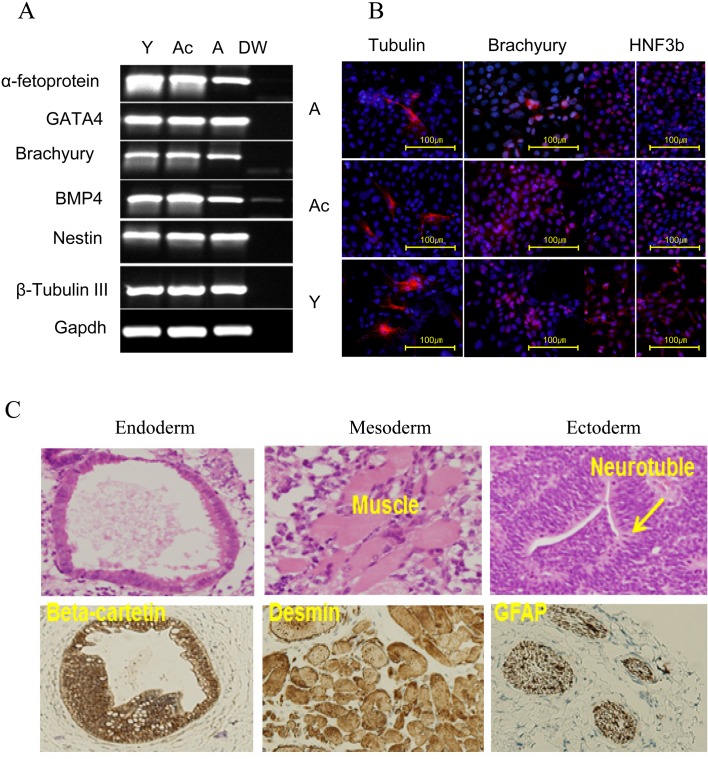
Fig. 6.
Differentiation of iPS cells in vitro and in vivo. A) RT-PCR analysis of differentiation markers for the three germ layers (endoderm, GATA4 and α-fetoprotein; mesoderm, brachyury and GATA2; and ectoderm, NESTIN and βIII-tubulin). Of note, a faint signal of BMP4 RT-PCR product in distilled water may be caused by flowing during application of electrophoresis to an RT-PCR product derived from A-derived iPS cells. Y, Ac, A and DW indicate Y-, Ac-, A-derived iPS cells, and distilled water, respectively. B) Immunocytochemical analysis of representative iPS cell lines during in vitro differentiation using three germ layer markers (ectoderm, tubulin; mesoderm, brachyury; endoderm, HNF3b). C) Teratoma formation analysis. Histological sections were examined with H&E staining and immunohistochemistry. Endodermal derivatives, epithelia, expressed a high level of beta-catenin. Mesodermal derivatives were exemplified by striated muscle and smooth muscle (middle). Tissues of ectodermal lineage, including the neural epithelium, expressed glial fibrillary acidic protein (GFAP).

