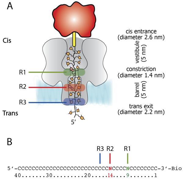
Figure 1.
DNA immobilized within the αHL nanopore. A) Cartoon representation of a homopolymeric DNA oligonucleotide immobilized inside an αHL pore (grey) through the use of a 3′ biotin-TEG (yellow)-streptavidin (red)complex. The green, red and blue boxes represent R1, R2 and R3 recognition sites, respectively. B) The sequences of the oligonucleotides with single nucleobase substitutions at position 9 (green) and 14 (red) that were used to probe R1 and R2, respectively. The recognition site R3 is shown as a blue line.

