
Figure 3. PC reduces SIRT1 levels in the injured lung.
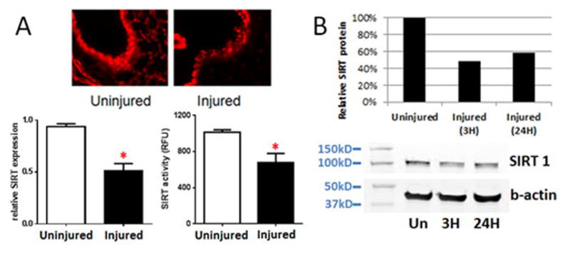
After 24 hours, lung tissue from injured mice was removed and compared to lung tissue from uninjured mice. Lung tissue (A) was fixed and stained for SIRT1 protein (upper panel) or processed for RNA analysis (lower left) or SIRT activity (lower right). A representative section of uninjured (n=1) and injured (n=3) lung is shown. PCR results shown are reported as relative SIRT expression using GAPDH mRNA as the internal control (n=8, *p<0.0001). SIRT activity results are reported as specific SIRT1 activity in RFU (n=3 uninjured; n=4 injured, *p<0.05). At 3 or 24 hours after PC, BAL cells from injured mice (n=4) were harvested, pooled and analyzed for SIRT1 protein (B). SIRT1 or b-actin (loading control) levels were quantitated by densitometric analysis of the immunoblot and reported as SIRT1 protein levels in injured mice relative to uninjured mice.