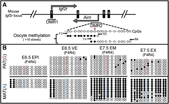
Figure 2.

DNA methylation at DMR2. (A) Schematic of the mouse Igf2r/Airn locus: transcription start sites (bent arrows), Igf2r exons (solid boxes), and location of DMR1 and DMR2. Two amplicons (F2R2 and F4R4) spanning 20 CpG dinucleotides were analyzed. Methylation in oocytes confirmed that DMR2 is an ICR and defined the methylation boundary (red asterisk in A). (B) ICR methylation is maintained in EPI and VE, but spreading of maternal DMR2 methylation occurs in EM and EX (compare EPI to EM, and VE to EX). Filled circles = methylated cytosine, Open = unmethylated cytosine. Asterisk denotes ICR border. Arrows indicate bisulfite-sequencing primer locations in (A). Parental SNP indicated on each bisulfite strand. Pat, paternal; Mat, maternal.
