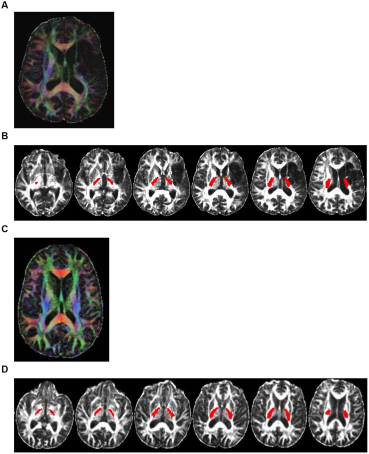
FIGURE 1.
Color-coded diffusion tensor imaging (DTI) images and the PLIC white-matter tract. Directionally encoded color (DEC) map of DTI images and the PLIC white-matter tract. (A) A DTI image of a representative patient (CI001) with damage to PLIC. Color codes to give diffusion tensor directions: red represents tracts running left to right; green is anterior to posterior; blue is superior to inferior. (B) Axial view of the PLIC white-matter tract (in red) of patient CI001. (C) A DEC map of DTI image of a representative patient (CI003) with no damage to PLIC. Color also represents diffusion tensor directions. (D) Axial view of the PLIC white-matter tract (in red) of patient CI003.

