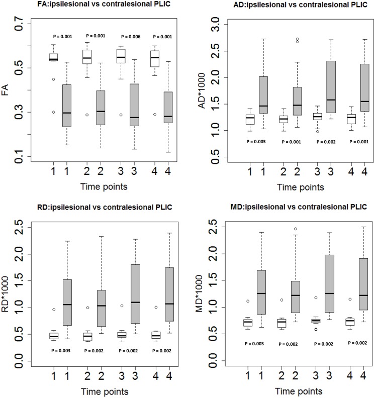
FIGURE 2.
Diffusion tensor imaging measures in ipsilesional and contralesional PLIC. Boxplots showed significantly higher FA, lower diffusivity measures (AD, RD, and MD) in the ipsilesional PLIC (Wilcoxon signed-rank tests).Time of immediately pre-, mid-, immediately post- and 1-month-post intervention are indicated as time-point 1, 2, 3, and 4, respectively. White boxes represent the contralesional side and gray boxes represent the ipsilesional side of PLIC.

