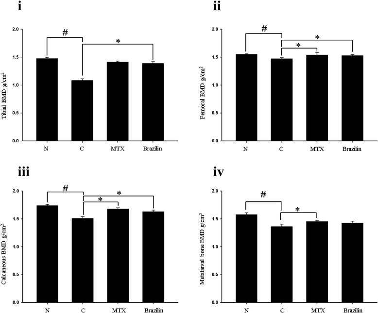
Figure 5.

Bone mineral density (BMD) of (i), the proximal part of the left tibial metaphysis; (ii), the distal part of the left femoral metaphysis; (iii), the distal part of the left calcaneous; and (iv), the distal part of the left second metatarsal bone. Values are means ± standard errors.*p < 0.05 (versus CIA mice); # p < 0.05 (versus normal group. Statistical analysis employed by Fisher’s protected least difference post hoc test.
