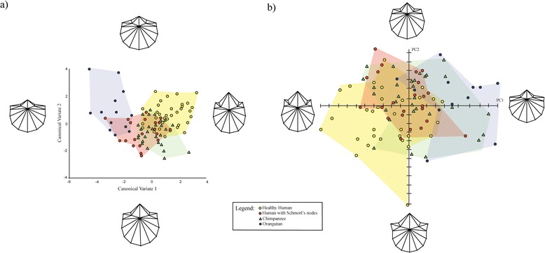
Figure 3.

CVA and PCA plots depicting shape variance of L1 vertebrae. a) CVA scatter-plot illustrating shape variation of healthy human, pathological human, P. troglodytes, P. pygmaeus vertebrae on CV1 and CV2 for L1 vertebrae b) PCA scatter-plot illustrating shape variance of healthy human, pathological human, P. troglodytes, P. pygmaeus vertebrae on PC1 and PC2 of L1 vertebrae. Deformation grids illustrate shape differences occurring on each PC.
