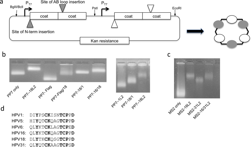
Figure 1.
Design and characterization of hybrid bacteriophage VLPs. A: Design of the hybrid VLP expression plasmid. Peptide targets can be displayed at either the N-terminus or the AB-loop of the coat protein single-chain dimer. Each expression cassette is engineered separately, amplified by PCR, and then plasmids are assembled by three-piece ligation using the restriction sites listed. B: PP7 or C: MS2 VLPs were analyzed using a 1% agarose, non-denaturing gel stained with ethidium bromide (which binds to the genomic material encapsidated by the VLPs). The mobilities of the bands were compared to VLPs of unmodified PP7 or MS2 coat protein. D: An alignment of selected HPV sequences representing L2 aa 17–31 (or the equivalent).

