
Figure 3.
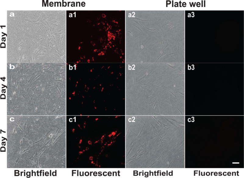
Transwell culture shows a lack of cross-labeling of human mesenchymal stem cells (hMSCs) by bioconjugated quantum dots (QDs). QD-labeled hMSCs were cultured in the insert of a transwell system. The diameter of the insert is 400 nm, much larger than the diameter of QDs in the range of 2–10 nm. Unlabeled hMSCs were cultured underneath in the transwell plate. Whereas QD-labeled hMSCs were observed under fluorescent microscope during the tested 1, 4, and 7 days (a1, b1, and c1), no apparent QD labeling was observed in the unlabeled hMSCs cultured underneath in the same medium (a3, b3, and c3). (a–c) Bright-field images of QD labeled hMSCs; (a2–c2) bright-field image of unlabeled hMSCs. These data suggest that QDs extruded by hMSCs are not taken up by the unlabeled hMSCs up to the tested 7 days of culture. Scale bar: 100 μm.