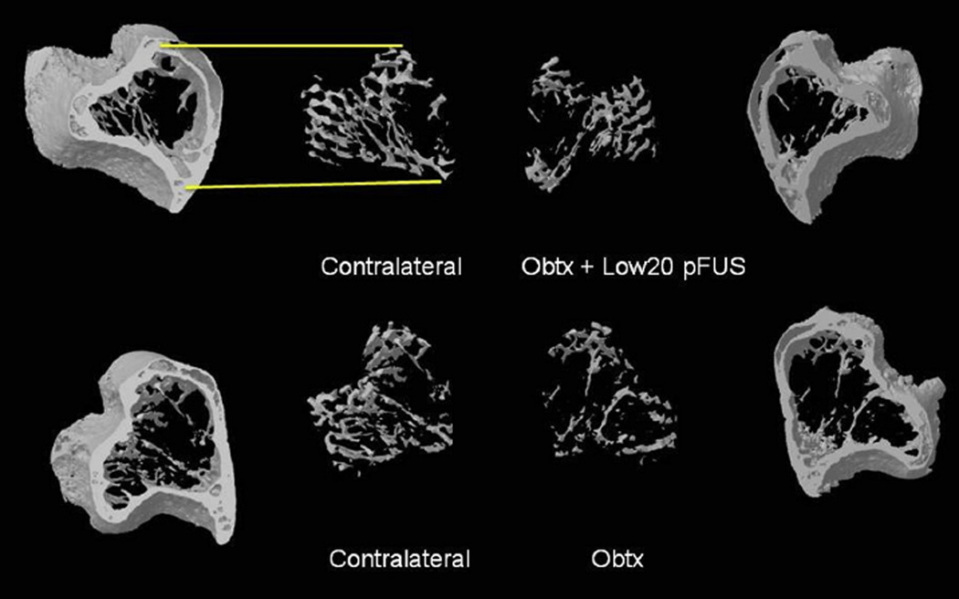
Fig. 5.
Example images of 3D volume renderings of micro-computed tomography data reveal the proximal tibia with the trabecular bone extracted in both the experimental (right) and contralateral (left) limbs. Top: Animal injected with ona-botulinumtoxinA (Obtx) and treated with pulsed focused ultrasound (pFUS) using the Low20 treatment protocol. Bottom: Animal from the Obtx sham group. This image qualitatively indicates the ability of pFUS treatment of the paralyzed muscle to mitigate paralysis-induced trabecular bone loss (Obtx + Low20 pFUS). Mean values for the different parameters characterizing bone loss for all experimental and sham groups are given in Figure 4.

