Fig. 6.
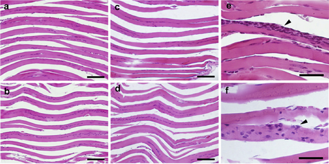
Representative histologic images of formalin-fixed calf muscle stained with H&E. Images reveal longitudinal muscle fibers (pink) with nuclei (purple). No damage was detected in the (a) Obtx sham, (b) Low20-treated, (c) High80-treated, or Saline sham (image not shown) limb compared with (d) an uninjected contralateral limb. There was some evidence of localized mild perifascicular inflammation in many of the injected tissue muscles, whether sham treated or ultrasound treated. Examples of inflammation {arrowhead) in Obtx sham (e) and High80-treated (f) samples are presented. Bar = 100 µm (a–d) and 40 µm (e–f).

