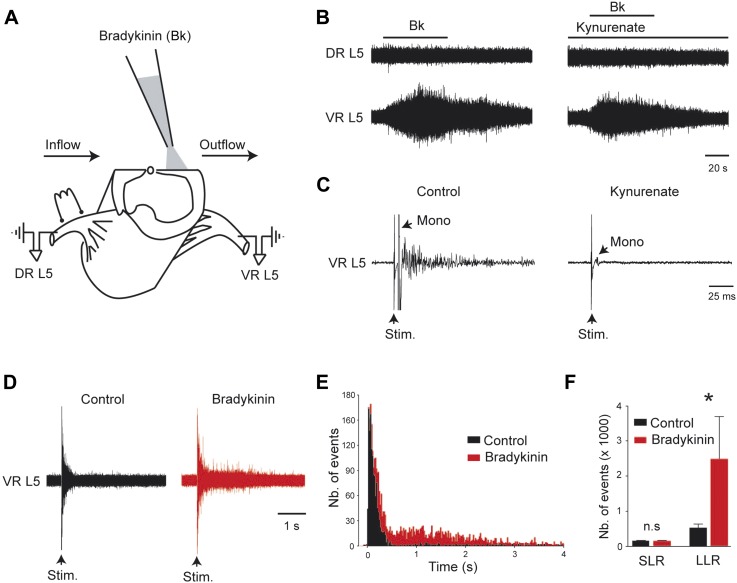
Figure 1. Bradykinin potentiates the gain of sensory inputs into the motor system.
(A) Drawing of a midsagittally hemisected rat spinal cord illustrating localized Bk application to the lumbar motor column, and dorsal (DR) and ventral (VR) roots used for reflex testing. (B) Responses to ventral application of bradykinin (Bk, 4 µM pipette concentration) recorded via the lumbar L5 dorsal (DR L5) and ventral (VR L5) roots before (left) and after (right) the fast glutamatergic synaptic transmission was blocked by kynurenate (1.5 mM). (C) Ventral root response to ipsilateral dorsal root stimulation before (left) and after (right) application of kynurenate (1.5 mM). Single arrows indicate the monosynaptic reflex (mono) and the stimulus artifact (stim). (D) Five superimposed responses recorded from an L5 ventral root induced by stimulations of the ipsilateral dorsal root before (control, black trace) and after local application of low concentrations of Bk (1 µM pipette concentration, red trace). (E) Average peristimulus time histogram (PSTH, bin width: 20 ms) of L5 ventral root recordings collected before (black) and after (red) the local application of Bk (1 µM pipette concentration). (F) Group means quantification of the PSTH for the transient short latency and long-lasting reflexes computed over a window 10–40 ms and 500–4000 ms post stimulus, respectively, before (black) and after (red) local application of Bk. Error bars indicate SEM. *p < 0.05 (Wilcoxon paired test).


