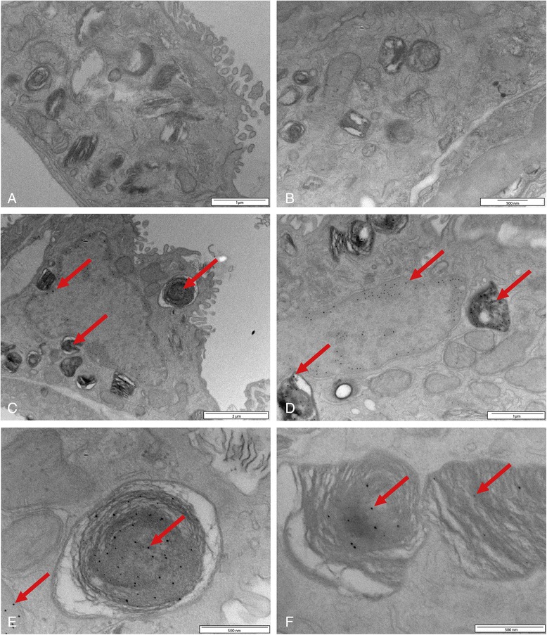
Figure 3.

TEM pictures of lung tissue of rats exposed to clean air (A and B) or 15 nm silver nanoparticles (C, D, E and F), afterwards followed by silver enhancement. The pictures show the presence of silver nanoparticles in the nucleus, lamellar bodies and lysosomes of lung cells (indicated by the red arrows) at 24 hours after exposure.
