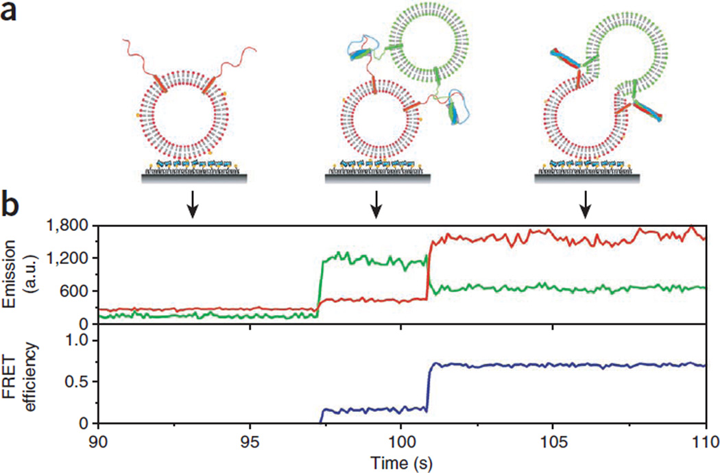
Figure 6.
Real-time lipid mixing for lipophilic dyes. (a) Cartoons showing different stages of fusion reaction between a vesicle pair. (b) Fluorescence intensity time traces of the donor (DiI, green) and the acceptor (DiD, red, top) and the corresponding trace of FRET efficiency, E (blue, bottom). No appreciable fluorescence signal change was observed until a t-SNARE liposome docked to a v-SNARE liposome. Rapid lipid mixing between the two vesicles caused by fusion leads to an increase in E. a.u., arbitrary units.

