
Figure 1.
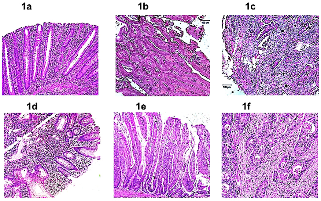
(a). Non-neoplastic Crohn disease. Section showing chronically inflamed colonic mucosa without architectural change (magnification, 100×). The cytological features of dysplasia are absent. (b). Crohn disease-associated (CD-a) dysplasia. Section of dysplasia in Crohn disease showing a tubulovillous adenoma-like dysplasia lesion (magnification, 100×). (c). Crohn disease-associated (CD-a) cancer. Invasive carcinoma in a Crohn disease patient showing infiltrating carcinoma glands set in tumor desmoplasia (magnification, 100×). (d). Non-neoplastic ulcerative colitis. Section showing chronically inflamed colonic mucosa without architectural change (magnification, 100×). The cytological features of dysplasia are absent. (e). Ulcerative colitis-associated (UC-a) dysplasia. High magnification view of part of a villous adenoma-like dysplastic lesion in a patient with ulcerative colitis (magnification, 200×). Note the full thickness stratification of nuclei with nuclei occupying the luminal portions on many of these colonic epithelial cells. (f). Ulcerative colitis-associated (UC-a) colorectal cancer. Infiltrating moderately differentiated adenocarcinoma characterized by neoplastic glands showing an infiltration pattern set in tumor desmoplasia (magnification, 200×).