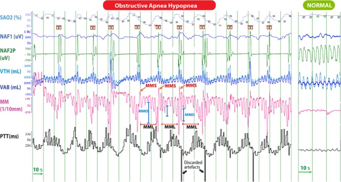
Figure 3A. Polysomnographic tracing of 4-min of stage N2 sleep, in a 12-year-old child with tonsillar hypertrophy, showing regular episodes of obstructive apnea-hypopnea.
When sampling the PTT signal at the same frequency as the MM signal and after adjustment for the lag of these signals, MM was highly correlated with PTT (Pearson r = 0.65; p < 0.001) in this patient; the more the mandible lowered during MMO, the more PTT lengthened. Cortical arousals are enclosed into the small brown symbols. On the right is a tracing during a quiescent period of normal respiration and little mandibular movement. SaO2, arterial oxyhemoglobin saturation; VTH and VAB; thoracic and abdominal inductance belts; NAF2P and NAF1, nasal pressure transducer and oronasal thermal flow sensor; MM, mandibular movements; PTT, pulse transit time; Phono, microphone sound; HR, heart rate; OAH, obstructive sleep apnea-hypopnea; MMS: sharp and sudden MM; MMC: peak to peak respiratory MM ≥ 0.3 mm; MMO: mouth opening.

