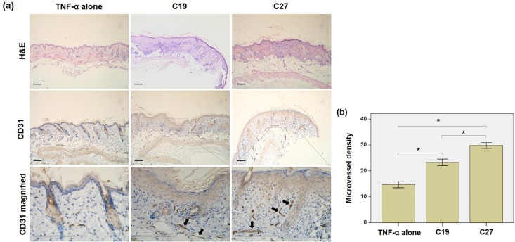
Fig 5. (a) Both C19 and C27 induced epidermis proliferation in BALB/c mice.
Mice were subcutaneously injected with C19 or C27 peptide. The paraffin sections were prepared from the skin tissues of injection sites. a By H&E staining, the peptide groups had significantly proliferated epidermis as compared to the control (TNF-α alone). Immunohistochemistry revealed stronger CD31 staining in dermal microvessels in the peptide groups as compared to the control. (b). The microvessel densities were quantitated in CD31 stained sections, showing significant increase in the peptide-injected mice especially in C27 group. Number of mice was five in each group. H&E, hematoxylin and eosin; TNF-α, tumor necrosis factor-α. Scale bar = 100 μm.

