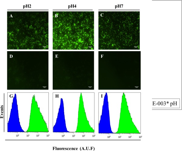
Fig 2. Detection of H. pylori in slides, by epifluorescent microscopy (A-F), and in suspension, by flow cytometry (H-J), using the FAM HP_ LNA/2OMe _PS oligonucleotides probe at different pH values.
A—F Smear of pure culture of H. pylori strain 26695 observed by epifluorescent microscopy. A-C. Experiment using 200 nM of the probe. D-F. Smears without probe were used as negative control. All images were taken at equal exposure times. G-I. Relative fluorescence histograms of LNA-FISH targeting H. pylori in different pH for two different assays—Blue: negative control with no probe; Green: positive sample. Scale bar = 10 μm.

