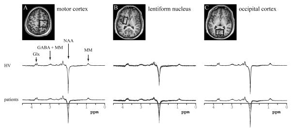
Figure 2. Regions of interest and data quality.
Upper part: The location and size of the voxels shown on T2-weighted images in the axial plane for (a) the motor cortex, (b) the lentiform nucleus, and (c) the occipital cortex.
Lower part: Data quality. All spectra obtained in healthy volunteers (upper row) and patients (lower row) in: (a) the motor cortex, (b) the lentiform nucleus, and (c) the occipital cortex. All spectra are shown with the vertical scale adjusted based on water reference. The edited MEGA-PRESS spectra allowed measurement of NAA, Glx and GABA concentrations. The resonances of each of those neurochemicals can be easily identified.
Abbreviations. NAA: N-acetylaspartate, Glx: glutamate + glutamine, GABA: γ-aminobutyric acid, MM: macromolecules.

