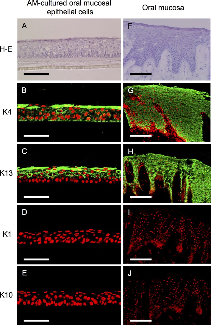
Fig 1. Morphology and keratin expression patterns of amniotic membrane-cultured oral mucosal epithelial cells and the oral mucosa.
Light micrographs of amniotic membrane (AM)-cultured oral mucosal epithelial cells and the in vivo oral mucosa stained with hematoxylin and eosin as well as representative immunohistochemical staining results of AM-cultured oral mucosal epithelial cells and the in vivo oral mucosa. Culture oral mucosal epithelial cells grew well on AM, exhibiting five to seven differentiated, stratified layers with a measurable thickness (A). Keratins 4 and 13 were expressed by AM-cultured oral mucosal epithelial cells (B and C). These keratins were expressed in all epithelial layers of the oral mucosa (G and H). Conversely, keratins 1 and 10 were not expressed in any layer of AM-cultured oral mucosal epithelial cells (D and E) or oral epithelial cells (I and J). Nuclei are stained with propidium iodide (red). Scale bars: (A–E) 100 μm, (F–J) 200 μm.

