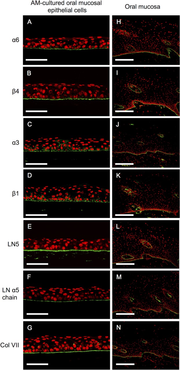
Fig 2. Expression of basement membrane proteins in amniotic membrane-cultured oral mucosal epithelial cells and the oral mucosa.
Representative immunohistochemical results. Positive staining for integrins alpha-6 beta-4 and alpha-3 beta-1, laminin 5, the laminin alpha 5 chain, and collagen VII was evident on the basement membrane side of the cultured oral mucosal epithelial cell layer (A–G) and the basement membrane of the oral mucosa (H–N). Nuclei are stained with propidium iodide (red). Scale bars: (A–G) 100 μm, (H–N) 200 μm.

