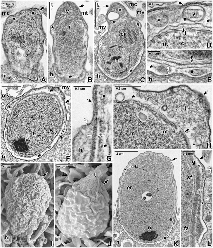
Fig 2. Ultrastructural features of Eleutheroschizon duboscqi early development.
A. Early trophozoite in the process of transformation from an attached zoite, already enveloped by a host-derived PS. TEM. B. Early trophozoite. Note the numerous vesicles in the space between parasite and PS, especially in caudal region. TEM. C-E. Young trophozoite. D shows the space between the parasite caudal region and PS; E shows the host parasite interface at the attachment site. TEM. F-H. Maturing trophozoite. G shows an annular joint point of two host membranes; H focuses on the parasite caudal region and PS. TEM. I. Early trophozoite. SEM. J. Young trophozoite. SEM. K. Mature trophozoite. TEM. L. Detailed view of the attachment site of the trophozoite shown in K, focusing on the developing fascicles of filaments and the annular joint point. TEM. a—parasite amylopectin, asterisk—space between the parasite and PS, black arrow—PS, black arrowhead—parasite plasma membrane, black double/paired arrowheads—parasite cytomembranes, c—parasite cytoplasm, cr—crystalloid body, er—parasite endoplasmic reticulum, fa—attachment fascicles, fi—short attachment filaments, g—glycocalyx, h—host cell, mc—host microcilia, mt—parasite subpellicular microtubules, mv—host microvilli, n—parasite nucleus, r—parasite posterior ring, sf—parasite subpellicular filaments, t—tail of the PS, v—vesicles, white arrow—host cell plasma membrane, white arrowhead—dense band, white double arrowhead—base of the PS (membrane of host cell origin), x—forming attachment fascicles.

