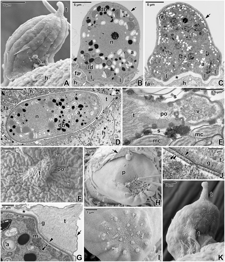
Fig 4. Morphology of Eleutheroschizon duboscqi gamonts.
A. Attached gamont. SEM. B. Macrogamont with a large central nucleus. TEM. C. Microgamont with several nuclei. TEM. D. Macrogamont enclosed by host tissue. TEM, RR. E. The PS tail of the macrogamont shown in D. Note the pores and the mucosubstances present in their surroundings. TEM, RR. F. High magnification of the caudal PS part with the tail showing numerous pores. SEM. G. Detailed view of the tail and gamont caudal part. TEM, RR. H. Upper view of an individual with a ruptured PS. SEM. I. The caudal region of a naked individual. SEM. J. High magnification of the interface between the parasite and PS in the area of the tail. TEM, RR. K. Gamont with two tails at the PS. SEM. a—parasite amylopectin, arrow—PS, asterisk—space between the parasite and the PS, black arrowhead—parasite plasma membrane, black double/paired arrowheads—parasite cytomembranes, c—parasite cytoplasm, db—parasite dense bodies, fa—attachment fascicles, g—glycocalyx, h—host cell, l—attachment lobe, ld—parasite lipid droplets, m—parasite mitochondria, mc—host microcilia, n—parasite nucleus, p—parasite, po—pore, s—mucosubstances, t—tail of the PS, v—vesicles, white arrowhead—base of the PS.

