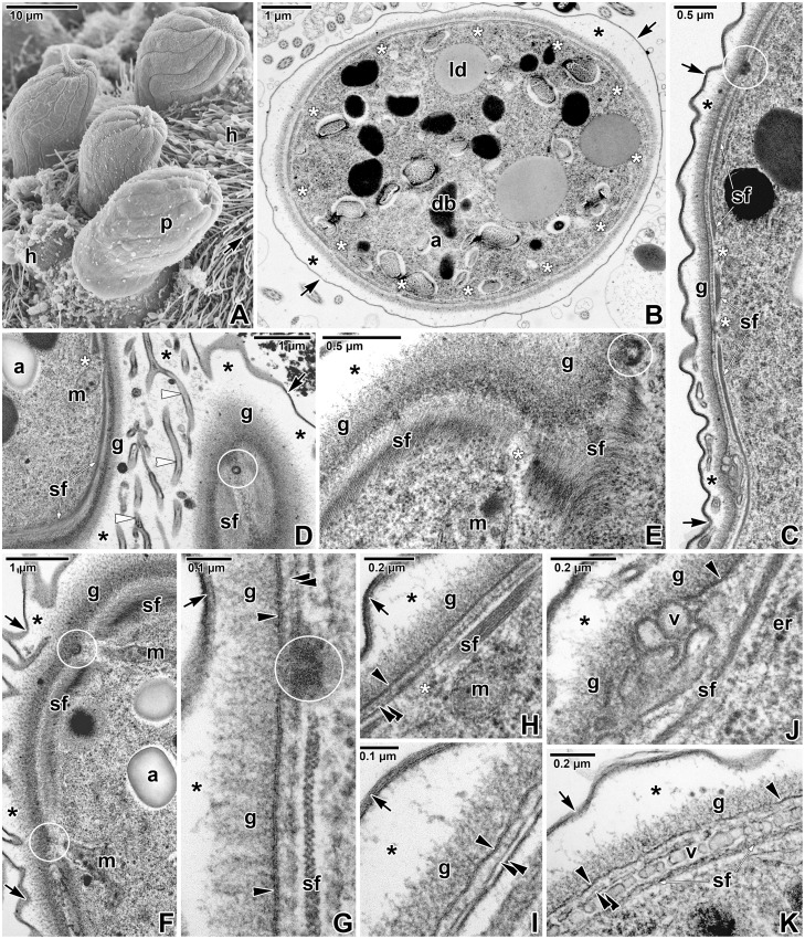
Fig 7. Fine structure of a parasitophorous sac and pellicle in Eleutheroschizon duboscqi gamonts.
A. Attached gamonts. SEM. B. Cross-sectioned gamont showing its surface organised in 12 broad folds and shallow grooves corresponding to the regularly arranged interruptions of subpellicular filaments. TEM. C. Longitudinal section showing the organisation of PS, gamont pellicle and the subpellicular layer of filaments that is repeatedly interrupted in areas corresponding to the localisation of micropores. TEM, RR. D. Superficial section of the PS and the gamont pellicle. The channel-like structures located in the space between the PS and parasite correspond to the folding of the PS observed under SEM. TEM, RR. E. Tangential section of the gamont surface underlined with subpellicular layer of filaments. TEM, RR. F. Diagonal section of the gamont surface revealing mitochondria connected with micropores. TEM, RR. G. Cross-section of pellicle showing the subpellicular filaments interrupted in the micropore area. TEM, RR. H. Almost longitudinal section of pellicle with interrupted subpellicular filaments. TEM, RR. I. Pellicle with continuous cytomembranes. TEM. J-K. Re-building of the parasite IMC indicated by the discontinuous cytomembranes and numerous vesicles located between the parasite plasma membrane and the subpellicular layer of the filaments. TEM, RR. a—parasite amylopectin, arrow—PS, asterisk—space between the parasite and PS, black arrowhead—parasite plasma membrane, black double/paired arrowheads—parasite cytomembranes, db—parasite dense bodies, er—parasite endoplasmic reticulum, g—glycocalyx, h—host tissue, ld—parasite lipid droplet, m—parasite mitochondria, p—parasite, sf—parasite subpellicular filaments, v—vesicles, white arrowheads—channel-like structures. Micropores are indicated by white circles, interruptions of subpellicular filaments—by white asterisks.

