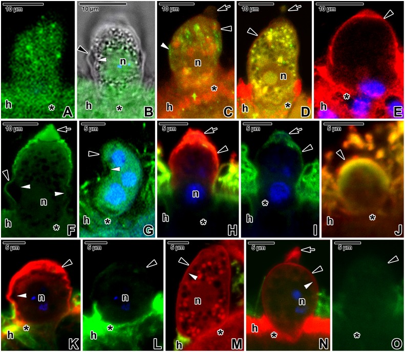
Fig 9. Immunolocalisation of Eleutheroschizon duboscqi cytoskeletal proteins.
A-B. Actin labelling with a medium intensity in a trophozoite (PFA fixation). CLSM, IFA (A) and CLSM in a combination with transmission LM, IFA/DAPI (B). B represents a single median optical section. C. Actin labelling in a macrogamont treated with 30 μM JAS for 7 hours (PFA fixation). Note the increased accumulation of parasite actin (FITC) organised in longitudinal bands exhibiting strong fluorescence and strong F-actin (TRITC) labelling with a diffuse character. CLSM, IFA/phalloidin-TRITC. D. A gamont exhibiting a more diffuse actin (FITC) labelling of medium intensity after treatment with 10 μM cytochalasin D for 9 hours (PFA fixation). The F-actin (TRITC) labelling of the parasite did not change significantly. CLSM, IFA/phalloidin-TRITC. E. Very strong myosin (TRITC) labelling restricted to the PS and host tissue (PFA fixation). CLSM, IFA/DAPI. F. Strong spectrin (FITC) labelling of the PS in a macrogamont (PFA fixation). CLSM, IFA/DAPI. Single median optical section. G. Labelling of α-tubulin (FITC) of strong intensity in a young microgamont (PFA fixation). CLSM, IFA/DAPI. H-I. A trophozoite (fixed in ice-cold methanol) exhibiting a labelling of medium intensity for α-tubulin (FITC) and very strong intensity for myosin (TRITC). CLSM, IFA/DAPI. J. Labelling of α-tubulin (FITC) and myosin (TRITC) in an early trophozoite treated for 7 hours with 10 μM oryzalin (fixed in ice-cold methanol). The fluorescence signals for both antibodies did not change significantly. CLSM, IFA. K-L. Localisation of α-tubulin (FITC) and myosin (TRITC) in an individual (probably a young microgamont) treated with 30 μM oryzalin for 3 hours (fixed in ice-cold methanol). The fluorescence signal for tubulin became very weak, while it remained very strong for myosin. CLSM, IFA/DAPI. M. Co-localisation of α-tubulin (FITC) and F-actin (TRITC) in a macrogamont treated for 7 hours with 10 μM oryzalin (PFA fixation). CLSM, IFA/phalloidin-TRITC. N-O. Labelling of α-tubulin (FITC) and F-actin (TRITC) in a maturing trophozoite treated for 3 hours with 30 μM oryzalin (PFA fixation). CLSM, IFA/phalloidin-TRITC/DAPI. In both the preparations (M-O), there was almost no fluorescence signal for α-tubulin, while the F-actin labelled with a strong intensity. arrow—tail of the PS, asterisk—parasite attachment site, black arrowhead—PS, h—host tissue, n—parasite nucleus, white arrowhead—parasite pellicle.

