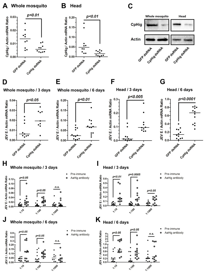
Fig 4. The antiviral effect of Hig in JEV infection of C. pipiens pallens.
(A-C) Inoculation of Culex pipiens pallens Hig (CpHig) dsRNA significantly decreased the CpHig expression in the whole mosquitoes and heads of C. pipiens pallens at both the mRNA (A and B) and protein (C) levels. The CpHig abundance was assessed by SYBR Green qPCR (A and B) and immuno-blotting with an AaHig antibody (C) at 6 days post dsRNA microinjection. (D-G) Silencing CpHig enhanced JEV infection in C. pipiens pallens. 10 M.I.D.50 JEV were inoculated at 3 days post CpHig dsRNA inoculation. The viral load of whole bodies (D and E) and heads (F and G) was assessed at 3 days and 6 days post-infection via Taqman qPCR and normalized with Culex actin. (H-K) Immuno-blockade of CpHig enhanced the JEV replication in C. pipiens pallens. The murine AaHig antibody, crossreacting with CpHig, was premixed with 10 M.I.D.50 JEV to co-inoculate into the Culex mosquitoes thorax. The treated mosquitoes were sacrificed to examine the viral load in the mosquito bodies (H and J) and heads (I and K) at 3 and 6 days post-infection by TaqMan qPCR and normalized against Culex actin. (D-K) The primers and probes of Taqman qPCR were described in the S1 Table. The experiments were repeated 3 times with similar results. One dot represents 1 mosquito and the horizontal line represents the median of the results. The data were statistically analyzed by the non-parametric Mann-Whitney test.

