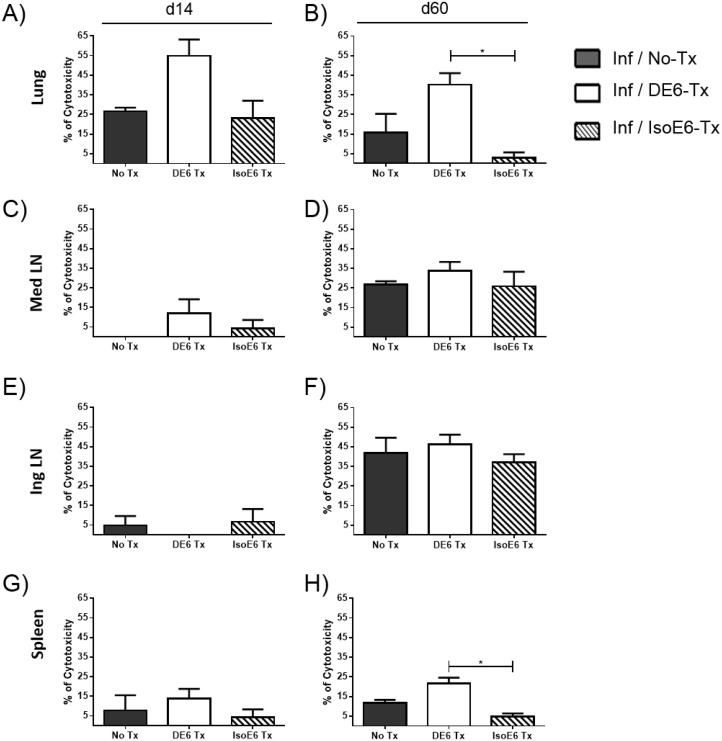
Fig 3. Lung in vivo target cell killing (CTL) rate in α-DEC-ESAT-treated mice is increased against ESAT p1 pool-loaded target cells.
The CTL activity was assessed in vivo at day 14 (left side panels: A, C, E, G) and day 60 (right side panels: B, D, F, H) after infection with virulent Mtb H37Rv. Two types of target cells were generated and stained differentially with CFSE alone or with CFSE plus PKH26. The target cells labeled with CFSE only were loaded with ESAT-6 pool 1 (p1) of peptides. Prior to transfer into different groups of Mtb-infected mice, both subsets of target cells were combined in equal proportions. The organs evaluated were the spleen, lungs, mediastinal and inguinal lymph nodes from mice treated with α-DEC-ESAT fusion antibody (Inf/DE6-Tx, white bars); and mice which received the isotype control antibody conjugated with ESAT-6 (Inf/IsoE6-Tx, stripped bars). Individual mice were analyzed and 3–5 mice were used per experimental group. Data are presented as mean plus standard error and percentage of cytotoxicity was calculated as indicated in Methods. (*) indicates P<0.05. All bars represent infected mice with different treatments for each group, as indicated.

