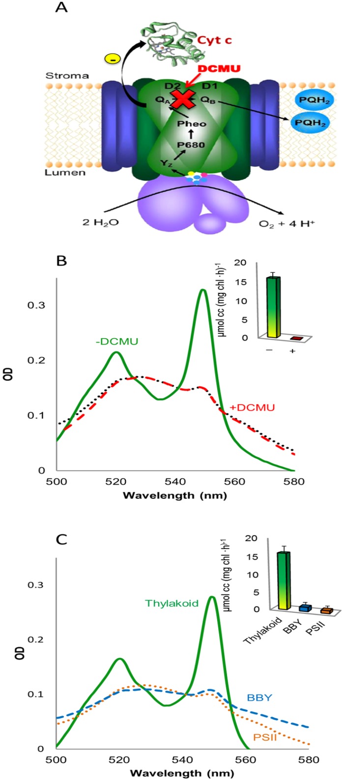
Fig 1. Cyt c is reduced by PSII in Syn but not in spinach thylakoids.

A. Schematic illustration of the postulated electron transfer pathway from PSII in Syn thylakoid to cyt cox. Photo-oxidized electrons are postulated to be transferred from QA to cyt c. The addition of DCMU blocks linear electron flow between the QA and QB and thus increases the reduction rate. B. Stacked spinach thylakoids were incubated for 3 min with cyt cox in the dark (black dotted), light (green) or in the light with the addition of DCMU (red dashed). Following centrifugation of the membranes, the absorption spectra of the cyt c containing supernatant was measured. The concentration of reduced cyt c was calculated using the coefficient Δϵ550nm-542nm. The inset depicts the quantification of cyt c photoreduction from three independent experiments in the absence (green) or presence of DCMU (red). C. Analysis of cyt c photoreduction at different levels of spinach PSII isolation. Unstacked spinach thylakoids (Thylakoid, solid green), PSII enriched membranes (BBY, dashed blue) or purified PSII (PSII, dotted brown) were incubated and analyzed as described for panel A. The inset depicts the quantification of cyt c photoreduction by the unstacked thylakoids (green bar) PSII enriched membranes (blue bar) or purified PSII (brown bar) from three independent experiments. No photoreduction occurred in the presence of DCMU.
