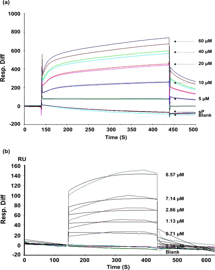
Fig 4. Sensogram showing binding of HbAHP-25 to gp120.
a) Different concentrations of HbAHP-25 and sP (20μM) were passed on gp120 immobilized chip. Direct binding was detected, represented as response units (RU). The image is representative of one of three identical experiments performed on three different days. b) Indicated concentrations of HbAHP-25 were passed over CM5 chip immobilized with gp120. Langmuir binding model (1:1) was used to evaluate binding constants. Sensograms are shown in different colors, corresponding to different concentration of HbAHP-25, and fits in black. Injections were carried in duplicates and results were identical.

