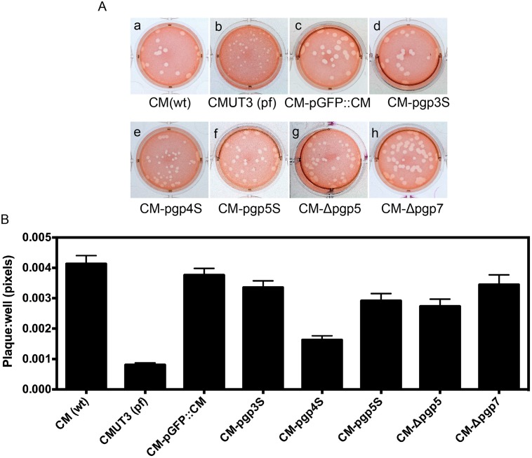
Fig 1. Comparison of plaque sizes among Chlamydia muridarum transformants.
(A) The following C. muridarum organisms were inoculated onto McCoy monolayers grown in 6-well plate: Wild type C. muridarum [CM(wt), image a], plasmid-free C. muridarum [CMUT3 (pf), b], CMUT3 transformed with the intact plasmid pGFP::CM (CM-pGFP::CM, c) or the pGFP::CM with a premature stop codon in pgp3 (CM-pgp3S, d), 4 (CM-pgp4S, e) or 5 (CM-pgp5S, f) or deletion of pgp5 (CM-Δpgp5, g) or 7 (CM-Δpgp7, h). The cultures were allowed to grow in 0.55% of argarose-containing medium for 5 days before neutral red staining and picture taking. (B) For quantitating the plaque sizes, the stained plates were scanned and the plaque sizes were measured in pixels using the customer-designed software PlaqueDetector. All plaque sizes were normalized using the areas of the corresponding wells so that plaque areas from different wells became comparable. The mean areas and standard deviations were used for comparing between different groups. Note that the plasmid-free CMUT3 and CM-pgp4S produced significantly smaller plaques (p<0.01, Student t-test).

