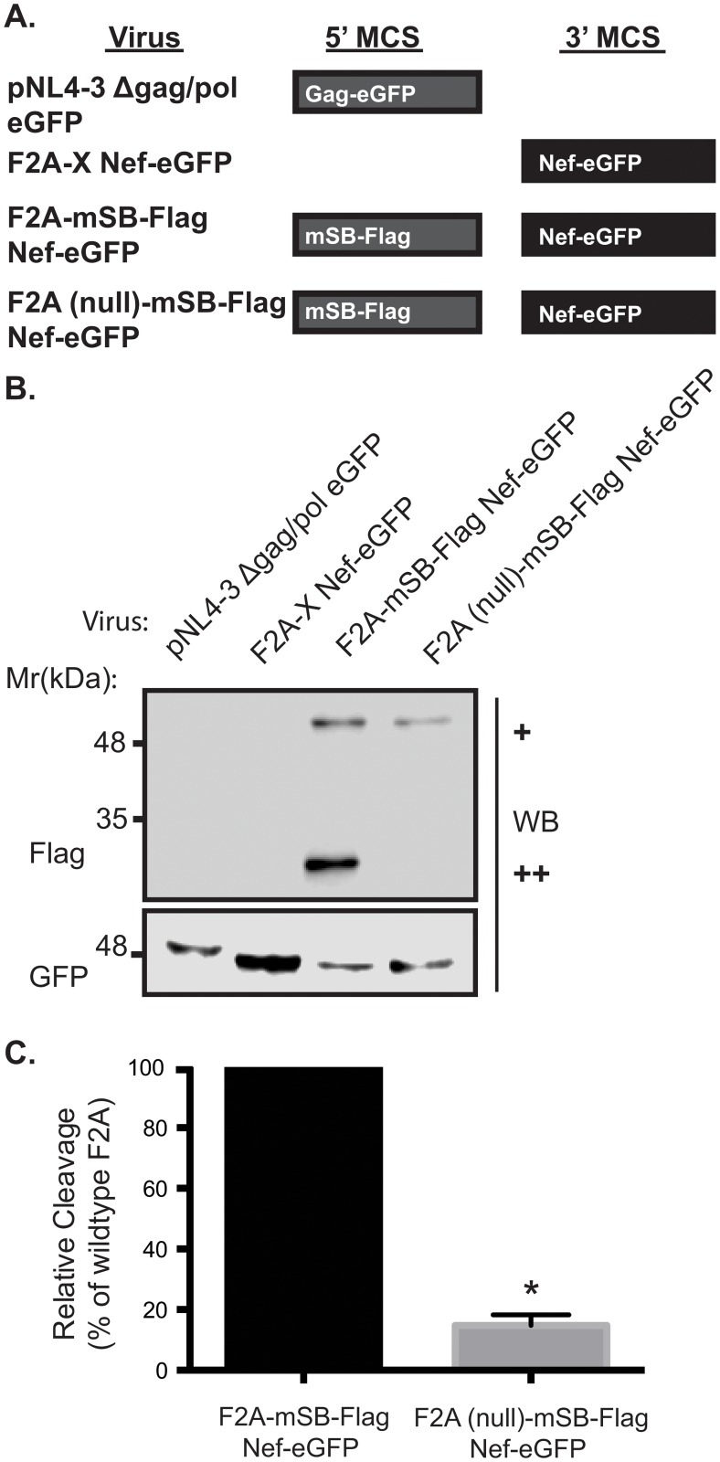
Fig 2. Functional cleavage at the engineered F2A site.
Viruses were engineered with various proteins within the 5’ MCS and/or the 3’ MCS and Jurkat E6.1 T-cells were infected with the resulting pseudoviruses. Flag and GFP specific western blots were performed on lysates collected 48 hours post infection to verify protein expression levels. (A) Schematic representation of proteins produced from lentiviral expression system. (B) A Flag specific western blot was used to quantitate the cleavage efficiency at the F2A site in the F2A-mSB-Flag Nef-eGFP virus, compared to the F2A mutant, F2A (null)-mSB-Flag Nef-eGFP, which lacks cleavage activity (+ uncleaved product, ++ cleaved product). GFP specific western blots confirmed the presence of the Gag-eGFP fusion protein (lane 1) or Nef-eGFP fusion proteins (lanes 2–4). (C) Cleavage efficiency at the F2A site was 6-fold higher compared to the F2A (null) virus (* indicates p-value < 0.05). Details on how the cleavage efficiency was calculated are included in Materials and Methods. Error bars calculated from 3 independent experiments. p-value was determined by paired t-test.

