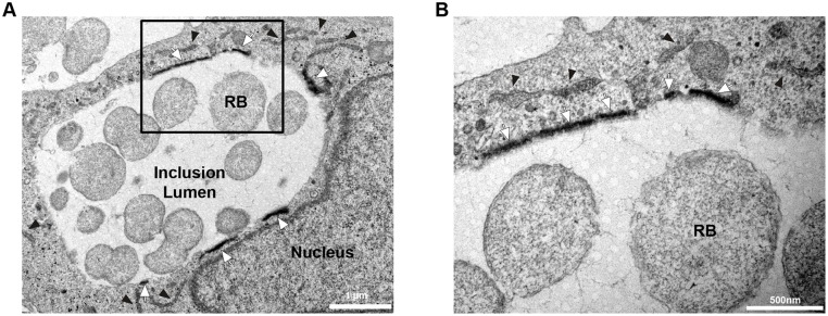
Fig 3. STIM1 is enriched at ER-Inclusion MCSs.
Electron micrographs of HeLa cells expressing HRP-STIM1, infected with C. trachomatis for 24h and processed for conventional transmission electron microscopy coupled with peroxidase cytochemistry. White arrows and black arrowheads respectively indicate ER structures displaying high and low level of HRP-STIM1. The area outline by the box in A is shown at higher magnification in B. RB: Reticulate Body. Scale Bars: 1 μm (A), 500 nm (B).

