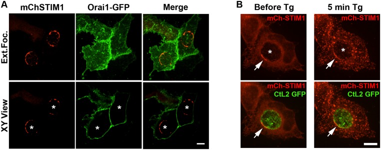
Fig 6. STIM1 cellular localization in response to Ca2+ store depletion.
A. Confocal micrographs of HeLa cells co-expressing mCherry-STIM1 (left panels, mChSTIM1, red) and Orai1-GFP (middle panels, Orai1-GFP, green) and infected with C. trachomatis for 24h. The merge is shown on the right. The top and bottom panels respectively correspond to the extended focus view combining all the confocal planes (Ext.Foc.) and a single plane crossing the middle of the inclusion (XY View). The asterisks in the XY View panels indicate the position of the inclusions. Scale bar: 10μm. B. Confocal micrographs of live HeLa cells expressing mCherry-STIM1 (mCh-STIM1, red) and infected for 24h with a strain of C. trachomatis that expresses GFP (CtL2 GFP, green). The images were acquired before (left panels, Before Tg) and 5 min after addition of Thapsigargin (Tg) (right panels, 5min Tg). The top and bottom panels respectively correspond to the mCh-STIM1 signal alone and the merge. An extended focus view combining all the confocal planes is shown. The arrow indicates STIM1 enrichment at ER-Inclusion MCSs. The asterisk indicates the position of the inclusion. Scale bar: 10μm.

