
FIGURE 1. Myocardial Infarct Models.
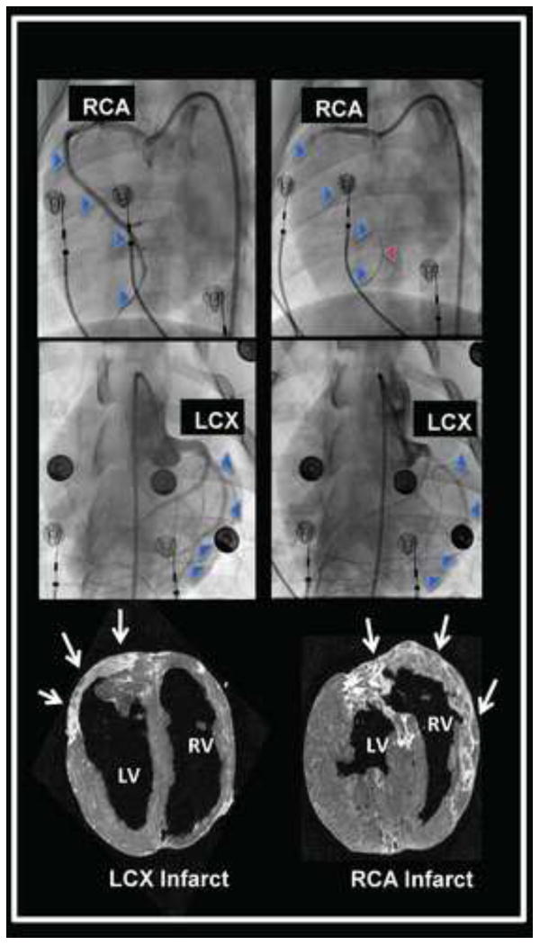
Angiographic evidence of occlusion of the right coronary artery (RCA) and left circumflex artery (LCX) to create right and left-sided myocardial infarctions are shown in the top and middle panels, respectively. The blue arrowheads trace the course of the vessel at baseline, and indicate the missing vessel following microsphere occlusion. The red arrowhead identifies an intracoronary guidewire tracing out the course of the occluded RCA. The bottom panel shows delayed gadolinium magnetic resonance images of the RCA and LCX infarcts (bright tissue), also identified by white arrows. Short axis images of the ventricles are presented, viewed from the apex of the heart.