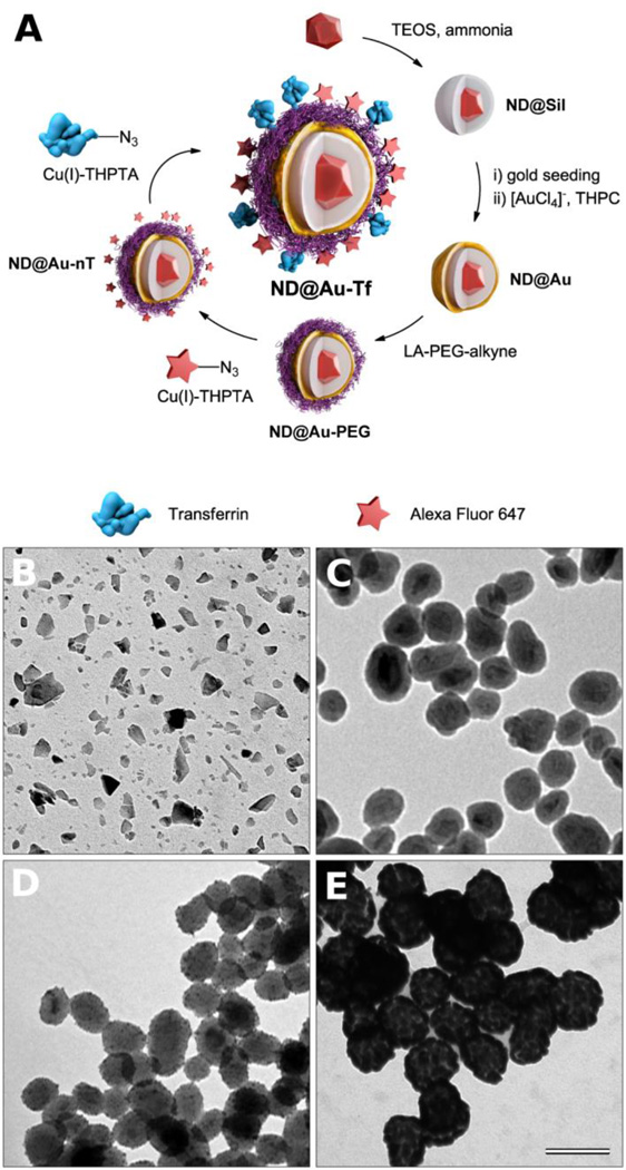
Figure 1.
(A) Schematic representation of the preparation of GNSs with a diamond core. First, a silica shell is created on diamond particles, followed by formation of a GNS upon reduction of [AuCl4]− promoted by adsorbed gold nanoparticle seeds. The GNS is modified with a lipoic acid-PEG conjugate, which is terminated with an alkyne. Using click chemistry, Alexa Fluor 647 dye and azide-modified transferrin (the targeting protein) are attached in consecutive steps. (B–D) TEM microphotographs of (B) diamond particles, (C) silica-coated diamond particles (ND@Sil), (D) silica-coated diamond particles with gold seeds, and (E) GNSs a with diamond core (ND@Au). The magnification is the same for all microphotographs, and the scale bar corresponds to 100 nm.

