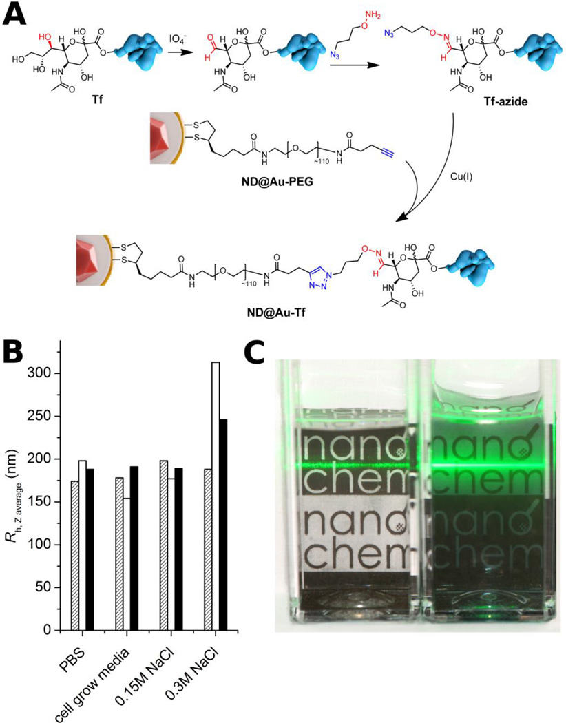
Figure 3.
Structure of ND@Au-PEG conjugate and its colloidal stability in aqueous solutions with high ionic strength. A) Composition of the particle surface architecture after modification and attachment of Tf. B) Hydrodynamic radii of ND@Au-PEG in various solutions after 1 h (hatched), 1 week (white) and 1 month (black), showing no aggregation. C) Photograph of naked (ND@Au, left) and PEG-coated (ND@Au-PEG, right) particles dispersed in PBS (20 min after mixing; 0.2 mg/mL concentration). The precipitating ND@Au particles are already partially sedimented on the bottom of the vial, while the remaining large aggregates unevenly scatter the laser beam. The ND@Au-PEG particles form a stable colloidal solution, which evenly and strongly scatters the laser beam.

