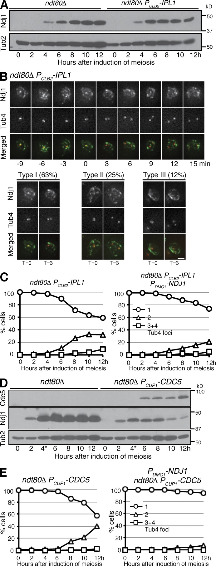
Figure 4.
Regulation of Ndj1 localization and protein stability during meiosis. (A) Immunoblot showing the Ndj1 protein level in ndt80Δ and ndt80Δ PCLB2-IPL1 (HY4506) cells. The level of Tub2 serves as a loading control. (B) Ndj1 localization in ndt80Δ PCLB2-IPL1 cells during meiosis. Time-lapse microscopy was performed as in Fig. 1 G. Red, Tub4-RFP; green, Ndj1-GFP. Bar, 2 µm. (C) SPB separation in ndt80Δ PCLB2-IPL1 and ndt80Δ PCLB2-IPL1 PDMC1-NDJ1 (HY4654) cells. SPBs were marked by Tub4-RFP. Note the delayed SPB separation in the presence of four copies of PDMC1-NDJ1 as shown in Fig. 3 E. The graphs shown are from a representative time-lapse experiment out of three repeats. (D) Immunoblot showing the Ndj1 protein level in ndt80Δ and ndt80Δ PCUP1-CDC5 (HY4074) cells. To induce CDC5 expression, 60 mM CuSO4 was added to the culture media 4 h (indicated by the asterisk) after induction of meiosis. (E) SPB separation in ndt80Δ PCUP1-CDC5 and ndt80Δ PCUP1-CDC5 PDMC1-NDJ1 (HY4803) cells during meiosis. The graphs shown are from a representative time-lapse experiment out of two repeats.

