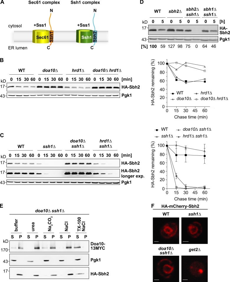
Figure 1.
Sbh2 is a Doa10 substrate and association with Ssh1 protects it from degradation. (A) Schematic of heterotrimeric yeast Sec61 and Ssh1 translocon complexes. The integral membrane protein Sss1, which is part of both complexes, is not depicted in the illustration. N, N terminus; C, C terminus. (B) Cycloheximide (chx) chase analysis of ectopically expressed (low-copy plasmid; MET25 promoter) HA-Sbh2 (in the presence of endogenous Sbh2) in WT, doa10Δ, hrd1Δ, and doa10Δ hrd1Δ cells. Pgk1 served as a loading control. The experiment shown is representative of n = 3 experiments. (right) Quantification of the gel on the left. HA-Sbh2 levels at t = 0 min were set to 100%. (C) Ssh1 protects Sbh2 from Doa10-dependent degradation. chx chase analysis of ectopically expressed HA-Sbh2 (as in B) in WT, ssh1Δ, doa10Δ ssh1Δ, and hrd1Δ ssh1Δ cells. Two different exposures of the anti-HA immunodetection are shown. The graph at right shows the mean degradation rates observed from three independent experiments. HA-Sbh2 levels at t = 0 h were set to 100%. Error bars represent ± SD. exp., exposure. (D) Degradation of unassembled Sbh2. chx chase analysis (time points t1 = 0 h and t2 = 5 h) of ectopically expressed HA-Sbh2 (as in B) in WT, sbh2Δ, sbh2Δ ssh1Δ, and sbh1Δ ssh1Δ cells. Relative protein levels listed below the blots were determined by quantification of pixel densities of HA-Sbh2 bands relative to those of Pgk1. HA-Sbh2 levels of WT cells at t1 = 0 h were set to 100%. (E) HA-Sbh2 is an integral membrane protein in ssh1Δ cells. Subcellular fractionation of doa10Δ ssh1Δ cells expressing HA-Sbh2 from a plasmid (as in B). HA-Sbh2–expressing ssh1Δ doa10Δ cells and Doa10-13MYC–expressing cells were mixed at a 5:1 ratio before lysis. Lysates were treated with buffer alone or buffer containing 2.5 M urea, 0.1 M Na2CO3, pH 11.5, and 0.5 M NaCl or 1% Triton X-100 (TX-100) and 0.5 M NaCl, and divided into microsomal pellet (P) and supernatant (S) fraction by centrifugation. Fractions were examined by immunoblotting with appropriate antibodies. (F) Fluorescence microscopy of WT, ssh1Δ, ssh1Δ doa10Δ, and get2Δ cells overexpressing HA-mCherry-Sbh2 from a low-copy plasmid under the strong TEF2 promoter. Bars, 1 µm.

