Figure 4.
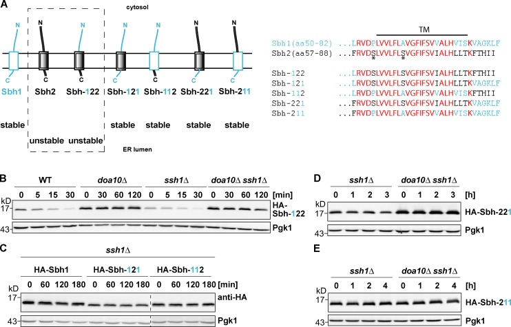
Investigation of Sbh1/Sbh2 chimeric proteins. Both Sbh2 TM and ER-luminal regions contribute to the degron. (A) Schematic representation of Sbh1, Sbh2, and Sbh1/Sbh2 chimeric proteins used in this study. Sbh1-derived regions are depicted in light blue, and Sbh2-derived regions are shown in black. The nomenclature for Sbh1/Sbh2 chimeric proteins is as follows: “Sbh-XYZ” designates a chimeric protein in which the number at position X indicates the source of the cytosolic domain (“1”: Sbh1; or “2”: Sbh2), the number at position Y indicates the source of the TM helix, and the number at position Z indicates the source of the ER-luminal domain, respectively. (right) Comparison of Sbh1, Sbh2, and Sbh1/Sbh2 chimera TA sequences. Residues depicted bold in red represent identical residues shared between Sbh1 and Sbh2; residues depicted in light blue are Sbh1-specific residues, and residues depicted in black indicate residues specific for Sbh2; the black horizontal line indicates the TM helix, and the asterisks indicate Sbh2 residues Ser61 and Ser68, respectively. N, N terminal; C, C terminal. (B) Degradation of HA–Sbh-122. chx chase as in Fig. 1 B. Note: There are different chase times for WT and ssh1Δ strains (30-min chase) and doa10Δ and doa10Δ ssh1Δ strains (120-min chase). (C) Degradation of HA-Sbh1, HA–Sbh-121, and HA–Sbh-112. All blots are from same experiment/gel but were cropped for clarity (dashed line). (D) Degradation of HA-Sbh-221. (E) Degradation of HA–Sbh-211.

