. 2015 Apr 23;15(2):100–109. doi: 10.4110/in.2015.15.2.100

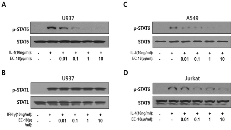
Figure 4. EC-18 dephosphorylated activated STAT6. Phosphorylation of STAT6 was examined using antiphospho STAT6 in the U937 (A), A549 (C), and Jurkat (D) cell lysates treated with EC-18 with concentrations from 0.01 to 10 µg/ml. Phosphorylation of STAT6 was induced by 10 ng/ml of the IL-4 treatment. (B) STAT1 phosphorylation was checked out in the cell lysate treated EC-18 with concentrations from 0.01 to 10 µg/ml. The activation of STAT1 was induced by 10 ng of the IFN-γ treatment in the U937 cells. Dephosphorylation of STAT1 and STAT6 was examined at 15 min after stimulation with IFN-γ and IL-4, respectively, in the EC-18 pretreated cells.

Figure 4