. 2015 Apr 23;15(2):73–82. doi: 10.4110/in.2015.15.2.73

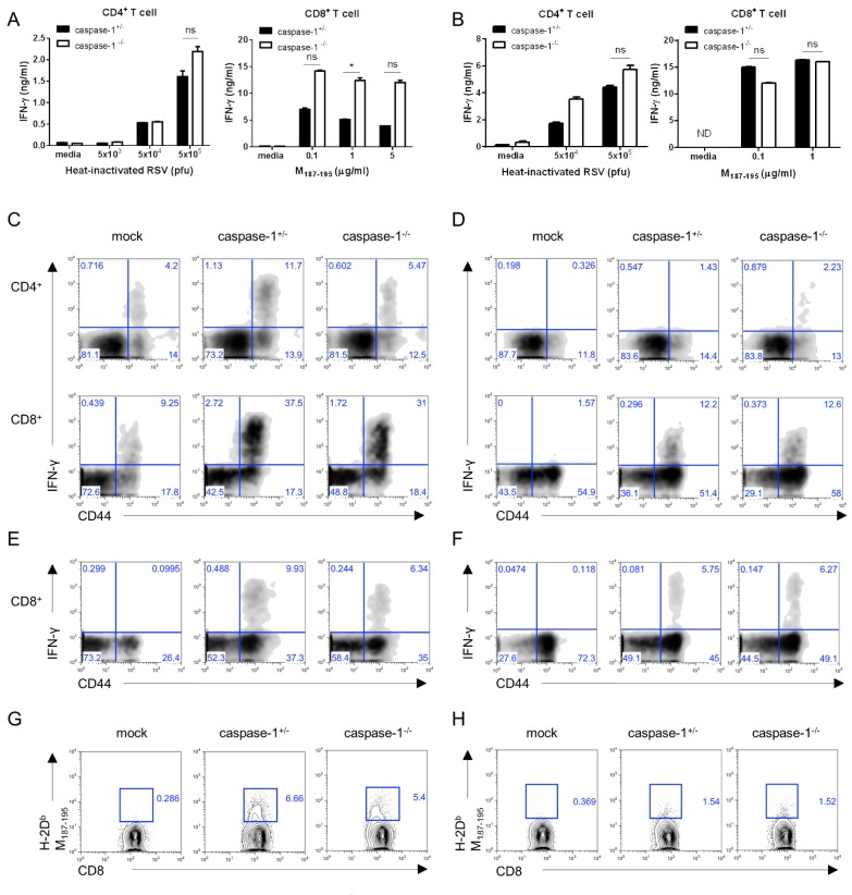
Figure 4. In 3-week- and 1-week-old mice, the caspase-1-/- mice T cell responses are normal. Three week (A, C, E, G) or one week (B, D, F, H) old caspase-1+/- and caspase-1-/- mice were intranasally infected with 1×10⁶ pfu of RSV. Caspase-1+/- mice were infected with PBS as a mock control (C~H). On day 8 after RSV infection, the mice were sacrificed. (A, B) CD4+ and CD8+ T cells from the mouse spleens were stimulated with the indicated heat-inactivated RSV or RSV M187-195 peptide, respectively, and pulsed with irradiated APCs. After 72 hr, IFN-γ in supernatants from the CD4+ T cells (left) and CD8+ T cells (right) were measured by ELISA. (C, D) IFN-γ-producing CD4+ T cells (up) and CD8+ T cells (down) were detected in the lungs by intracellular staining after re-stimulation with PMA and ionomycin or (E, F) RSV M187-195 peptide. (G, H) RSV-specific CD8+ T cells were stained with RSV tetramer (H-2Db M187-195) (G: 3-week, H: 1-week). Data are representative of two to three independent experiments. Error bars indicate SD.

Figure 4