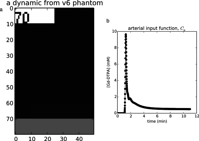
Figure 1. Example validation data.
(A) The noise-free v6 QIBA phantom contains a numeric label of its order in the time series in the upper left, the tissues regions in the middle, and a vessel strip at the bottom, from which the AIF may be extracted. (B) The AIF extracted from this phantom. (A) The noise-free v6 QIBA phantom contains a numeric label of its order in the time series in the upper left, the tissues regions in the middle, and a vessel strip at the bottom, from which the AIF may be extracted. (B) The AIF extracted from this phantom.

