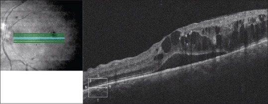
Figure 6.

Optical coherence tomography of the macula of the left eye of a patient with diabetic macular edema. There is subretinal fluid as well as cystoid intraretinal fluid present, causing loss of the normal foveal contour. There are areas of increased hyperreflectivity in the outer plexiform layer corresponding to hard exudates
