Figure 1.

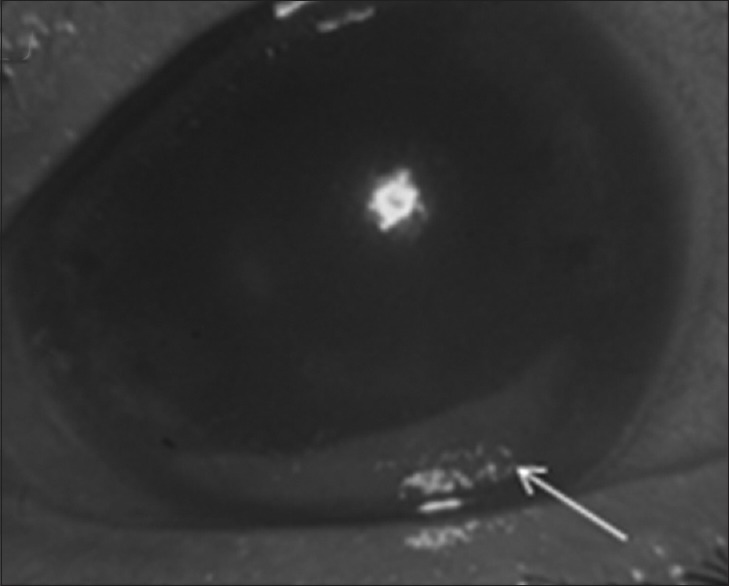
Anterior segment photograph of the left eye at presentation reveals diffuse corneal edema and a thick brown hypopyon (arrow)

Anterior segment photograph of the left eye at presentation reveals diffuse corneal edema and a thick brown hypopyon (arrow)