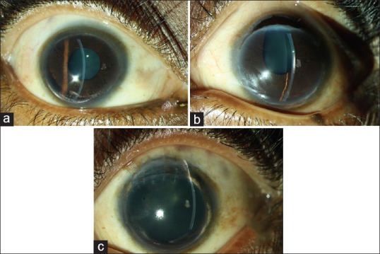
Figure 1.

(a) Slit lamp photograph of the right eye showing 360° peripheral corneal thinning with areas of lipid deposition and superficial vascularization. (b) Slit lamp photograph of the left eye showing a corneal perforation with iris prolapse adjacent to the limbus. (c) Slit lamp photograph of the left eye showing the clear graft superiorly with areas of lipid deposition superonasally and inferiorly
